
11
Few medical appointments provoke as much quiet apprehension as a breast imaging exam. Even when you schedule your annual appointment with routine nonchalance, receiving a follow-up call recommending an additional scan can make your stomach drop. For many women, that follow-up recommendation involves an ultrasound, sparking an immediate flurry of questions: Did the mammogram miss something? Is an ultrasound more accurate? Could I have simply skipped the compression plates and opted for an ultrasound from the start?
The confusion surrounding breast imaging is entirely understandable. Both 3D mammograms and breast ultrasounds are widely used, highly effective tools, yet they operate on fundamentally different scientific principles and serve distinct diagnostic purposes. One does not render the other obsolete. In reality, choosing the right screening is rarely a matter of picking one over the other; it is about understanding how these complementary technologies work together to provide a clear, detailed picture of your specific tissue composition.
Understanding 3D Mammography: Digital Breast Tomosynthesis
For decades, the standard 2D digital mammogram was the undisputed cornerstone of early breast cancer detection. While 2D imaging saved countless lives, it had a major physical limitation: it compressed three-dimensional breast tissue into two flat, overlapping images taken from the top and the side. For women with complex or fibrous tissue, that overlap could easily obscure small tumors or create false alarms that required stressful callbacks.
Modern 3D mammography, clinically known as digital breast tomosynthesis, was engineered specifically to solve the overlapping-tissue dilemma.
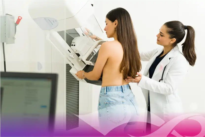
During a 3D mammogram, an X-ray arm sweeps in a gentle arc over the breast while it remains under light compression. In a matter of seconds, the machine captures a rapid sequence of low-dose images from multiple angles. Advanced computer software then synthesizes these exposures into a series of razor-thin cross-sectional slices, typically one millimeter thick.
A interpreting radiologist can scroll through these digital slices one layer at a time, much like turning the pages of an illustrated book. By visually peeling back the outer layers of tissue, the physician can inspect the internal architecture of the breast without distortion, spotting abnormalities that would otherwise remain hidden behind normal glandular structures.
The Defining Strengths of 3D Mammograms
The greatest clinical asset of 3D mammography is its ability to detect microcalcifications. These tiny specks of calcium, often smaller than a grain of salt, are frequently the earliest visible sign of developing breast cancer, particularly ductal carcinoma in situ. Ultrasound waves cannot reliably resolve microcalcifications, making mammography the only imaging tool capable of tracking these microscopic warnings before a physical mass ever forms.
Furthermore, 3D mammograms excel at providing a comprehensive, wide-angle overview of both breasts simultaneously. This architectural perspective allows radiologists to compare symmetry between the left and right breasts and monitor subtle architectural distortions over successive years.
How Breast Ultrasound Operates
Unlike mammography, which uses low doses of ionizing radiation, breast ultrasound relies entirely on high-frequency sound waves. A trained technologist applies a water-based gel to the skin and glides a handheld wand, called a transducer, across the surface of the breast.
The transducer sends sound waves deep into the tissue. As those waves encounter different structures, they bounce back at varying speeds. The system captures those returning acoustic echoes and translates them in real time into visual patterns on a monitor.
Ultrasound involves zero radiation exposure and requires no mechanical compression plates. However, its true value lies in how sound waves interact with soft tissue and liquid.
Differentiating Fluid-Filled Cysts from Solid Masses
The primary clinical superpower of an ultrasound is its ability to determine the internal composition of a lump. Sound waves pass straight through simple liquid without generating echoes, causing benign, fluid-filled cysts to appear as crisp, black, empty circles on the screen.
When sound waves hit a solid lesion, however, they bounce back with complex echoes, indicating that the mass consists of cellular tissue rather than fluid. Because millions of women naturally develop harmless, fluctuating cysts throughout their reproductive lives, an ultrasound can instantly resolve diagnostic panic by confirming that a palpable lump is merely a benign fluid pocket that requires no biopsy.
Evaluating Vascular Flow with Doppler Technology
Many modern ultrasound systems feature color Doppler imaging, which measures blood flow within and around a detected mass. Rapid, chaotic blood vessel growth is a common hallmark of malignant tumors, which build dedicated capillary networks to feed their expansion. By visualizing the presence and direction of blood flow, an ultrasound gives radiologists immediate physiological clues regarding whether a solid lesion behaves benignly or aggressively.
The Critical Variable: Breast Density
You cannot meaningfully compare mammography and ultrasound without addressing breast density. In recent years, healthcare providers and federal regulations have placed renewed emphasis on educating women about their individual density classification, and for good reason.
Breasts are composed of a mix of fatty tissue and dense fibroglandular tissue. On a mammogram, fatty tissue appears translucent and dark gray, creating high visual contrast. Dense glandular and connective tissue, however, appears solid white. The clinical challenge is that cancerous tumors and benign fibroadenomas also appear bright white on an X-ray.
When a woman has dense breasts, looking for a developing tumor on a mammogram is frequently compared to trying to spot a snowball in a blizzard. While 3D tomosynthesis significantly improves visibility compared to older 2D technology by isolating thin tissue slices, very dense tissue can still camouflage small abnormalities.
This is precisely where supplemental ultrasound screening becomes invaluable. Sound waves do not view dense tissue the same way X-rays do. While dense fibroglandular tissue shows up as bright white on a mammogram, it displays distinct grayish textural patterns under ultrasound, against which a dark, hypoechoic solid tumor stands out clearly. For women categorized with heterogeneously dense or extremely dense breasts, adding an ultrasound dramatically increases overall cancer detection rates.
Knowing When You Need a 3D Mammogram
For the vast majority of women, a 3D mammogram remains the non-negotiable frontline screening tool. You should prioritize a 3D mammogram under the following circumstances:
-
Your baseline annual screening: Beginning between ages forty and forty-five for average-risk women, an annual 3D mammogram provides the broadest, most reliable baseline view of long-term breast health.
-
Tracking structural symmetry over time: Mammograms are systematically cataloged and compared from year to year, allowing specialists to flag miniscule changes in tissue patterns before physical changes emerge.
-
Screening for asymptomatic microcalcifications: If you have no symptoms, no palpable lumps, and average breast density, a 3D mammogram provides the exact balance of comprehensive coverage and early-stage calcification detection you need.
Knowing When an Ultrasound Is Recommended
An ultrasound is rarely the first scan ordered for routine screening in the general population, but it becomes indispensable under specific diagnostic and supplemental conditions:
-
Investigating a palpable lump: If you or your physician feels a distinct lump, focal thickening, or localized pain during a physical exam, an ultrasound is typically ordered immediately to evaluate that specific geographic coordinate.
-
Evaluating dense breast notifications: If your mammogram report indicates that you have dense breast tissue, your physician will likely recommend a supplemental bilateral automated or hand-held screening ultrasound.
-
Clarifying ambiguous mammogram results: If a screening mammogram identifies an asymmetric shadow, a spot compression or targeted ultrasound is performed as a diagnostic workup to confirm whether the shadow represents real tissue or overlapping artifact.
-
Screening younger or pregnant individuals: For women under thirty who present with breast symptoms, as well as pregnant or nursing mothers, ultrasound is the preferred starting modality because it avoids radiation exposure entirely.
Why Ultrasound Cannot Simply Replace the Mammogram
Given that ultrasound involves no compression, emits no radiation, and can distinguish solid masses from fluid, many women naturally wonder why it cannot replace mammograms altogether.
The answer lies in screening limitations. Ultrasound is an exceptionally focused, operator-dependent examination. Unlike a mammogram, which captures standard, reproducible views of the entire breast in seconds, a handheld ultrasound requires a technologist to manually scan every square centimeter of tissue. Small lesions can be missed if the probe angle is slightly off.
More importantly, as noted earlier, ultrasound cannot reliably image the microcalcifications that signal precancerous cell changes. Relying solely on ultrasound would mean allowing non-invasive cancers to progress until they form physical, palpable masses before they could be seen.
Furthermore, ultrasound produces a substantially higher rate of false positives than mammography. It picks up countless benign abnormalities—such as tiny, harmless cysts, fibrous scar tissue, and normal lobules—that can mimic suspicious lesions on screen. This frequently leads to unnecessary biopsies, emotional distress, and costly follow-up procedures for lesions that were never dangerous to begin with.
Building Your Personalized Screening Strategy
Navigating breast health is not a one-size-fits-all equation. The most protective screening approach is personal, rooted in an accurate assessment of your family history, genetic background, and tissue density.
When you receive your imaging results, read beyond the simple clearance notice. Look for your breast density score, which will fall into one of four categories ranging from predominantly fatty to extremely dense. If your results show dense tissue, schedule a conversation with your healthcare provider about adding supplemental automated whole-breast ultrasound or handheld scanning to your annual routine.
By viewing 3D mammography as your broad, micro-level surveillance system and ultrasound as your targeted, depth-defining specialist, you can navigate your healthcare appointments with clarity, confidence, and the exact imaging your body requires.
